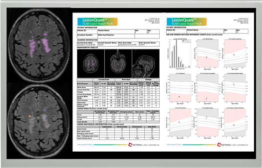
 Assessment of disease activity on MRIs from new and enlarging T2 FLAIR lesions and whole and regional brain volumes is an integral component of Multiple Sclerosis (MS) management.
Visit us at Stand A04 at the ECTRIMS – ACTRIMS Joint Meeting in Paris, October 25-28, to learn more about how LesionQuant can provide supportive information to physicians to aid in their clinical treatment planning and disease progression monitoring of patients with white matter diseases, such as Multiple Sclerosis.
The LesionQuant module of NeuroQuant (FDA cleared and CE marked) delivers fast, accurate anatomically color-coded segmented lesion images and automated FLAIR lesion and brain volume measurements compared to normative reference data. Additionally, lesion change visualization is available for review, when a prior study is provided.
Below are some recent blog posts which go into additional detail about how physicians can use quantitative imaging as biomarkers to evaluate lesion and brain structure volumes in patients with MS, and track longitudinal trends.
LesionQuant White Paper: Accuracy and Reproducibility of T2 FLAIR Segmentation
Using NeuroQuant in the Evaluation of Chronic Neurodegeneration
Mitigating the Risk of Repeated Gadolinium Use with LesionQuant
Defining and Classifying FLAIR Lesions in LesionQuant
Watch our Latest Webinar: An Introduction to LesionQuant
